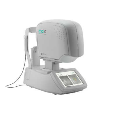
iCare MAIA
iCare MAIAiCare MAIA and S-MAIA confocal microperimetersKey features*25 Hz eye-tracking with confocal SLO imaging*Pre-programmed and custom tests*Sensitivity and fixation indexes, illustrative sensiti
- Weight::
- Brand ::
- No.::
iCare MAIAiCare MAIA and S-MAIA confocal microperimetersKey features*25 Hz eye-tracking with confocal SLO imaging*Pre-programmed and custom tests*Sensitivity and fixation indexes, illustrative sensiti
iCare MAIA and S-MAIA confocal microperimeters
*25 Hz eye-tracking with confocal SLO imaging
*Pre-programmed and custom tests
*Sensitivity and fixation indexes, illustrative sensitivity- and fixation maps
*Informative examination and progression reports
*2.5 mm minimum pupil size
*Patients with cataract (up to grade 3+) and media opacities can be examined
*Auto-focus (from -15D to +10D)
*Sensitive to functional changes due to macular pathologies or treatments even in early stages (background down to<0.0001 asb, threshold range 36 dB)
*Simple to use, patients can be tested in less than 3 minutes per eye
*S-MAIA includes also scotopic testing with cyan and red stimuli
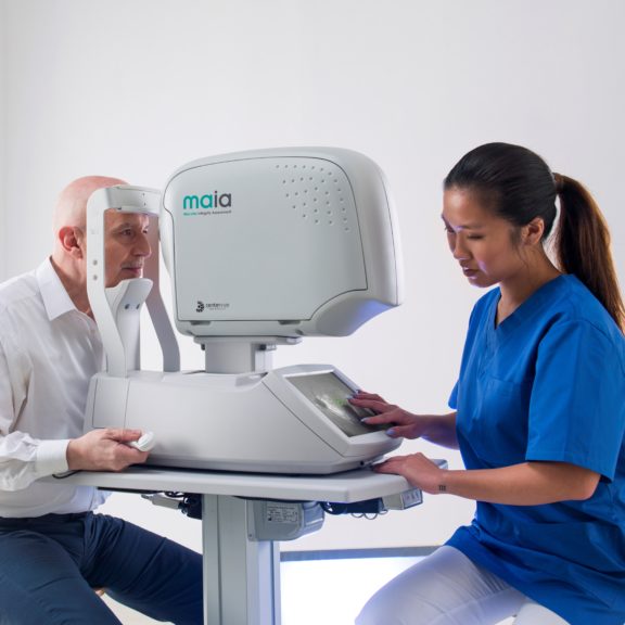
iCare MAIA (Macular Integrity Assessment) and S-MAIA offer the best in confocal microperimetry to combine visual field tests, fixation loss correction by a real-time retinal tracker and non-mydriatic confocal SLO fundus imaging, all in one exam. iCare MAIA and S-MAIA detect and monitor functional changes of the retina with great reliability.
iCare MAIA and S-MAIA perform different types of pre-programmed as well as customizable microperimetry tests and follow-up to monitor functional progression. The retinal tracker allows for accurate, real-time, calculation and compensation for eye movements. iCare MAIA and S-MAIA are patient friendly, automated, easy to use and non-mydriatic.
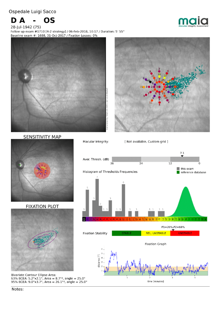
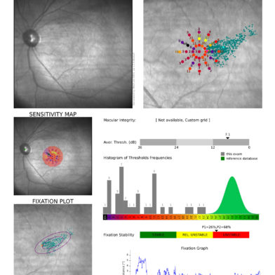
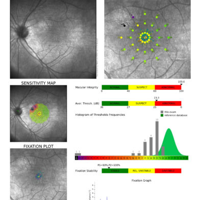
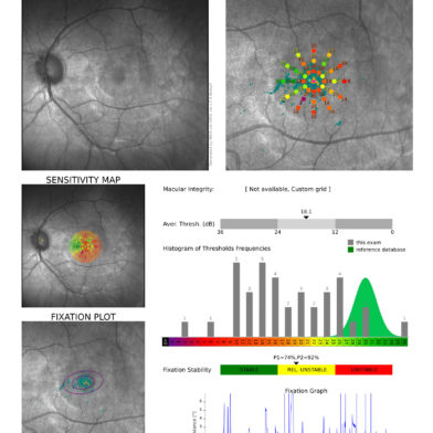
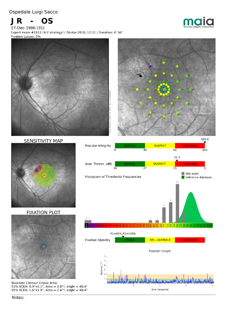
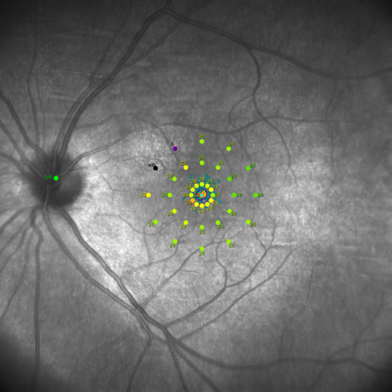
Microperimetry is a technology that allows concurrent analysis of structural and functional aspects of the retina. It combines retinal sensitivity mapping and fixation analysis with the fundus image in one exam as a powerful tool to detect, describe and follow-up pathologies affecting the macular area. iCare MAIA performs different types of microperimetry tests with supra and full-threshold strategies and follow-up tests to monitor functional progression. Each exam provides a measure of retinal sensitivity and fixation analysis (stability and position of the Preferred Retinal Locus).
Fixation analysis for a better diagnosis and vision
iCare MAIA provides accurate and objective information regarding retinal location and stability of a patient’s fixation. Parameters are assessed by tracking eye movements 25 times per second and by plotting the resulting distribution over the SLO image. iCare MAIA can project multiple fixation targets at selectable locations to help patients visualize the goal during the sensitivity test. The fixation test can also be done alone and takes only seconds. A fixation test can also be conducted to train the patient to use a new optimal Preferred Retinal Locus.
iCare MAIA performs different types of pre-programmed as well as customizable microperimetry tests and follow-up to monitor functional progression. The retinal tracker allows for accurate, real-time, calculation and compensation of eye movements.